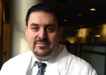
Joseph Sakran, associate professor of surgery, acute care, and trauma surgery, is a trauma surgeon, coalition builder, policy advisor, public health expert, and nationally recognized advocate for gun violence prevention. A survivor of gun violence, Dr. Sakran’s interest in medicine and trauma surgery began after a stray bullet nearly killed him during his senior year of high school. He has subsequently dedicated his life to treating the most vulnerable, reducing health disparities among marginalized populations, and advancing public policy that alleviates structural violence in low-income communities.
Dr. Sakran’s research focuses on firearm-related injuries with the goal of reshaping the cultural narrative around responsible gun ownership. Through in-depth analysis, qualitative assessments, and effective communication strategies, the objective is to collaborate with partners to instigate a shift in consciousness, preventing firearm-related injuries. The initiative involves a long-term public safety movement, aiming to foster behavioral shifts through strategic partnerships in healthcare, schools, and the media. Through a provocative social norm campaign, in collaboration with the Ad Council and PR agencies, Dr. Sakran aims to deliver compelling messages through creative ad campaigns. The final component of his plan includes thought leadership for future advocacy, cultivating support for future political advocacy, and testing policy messages.
Vishal Patel, associate professor of electrical and computer engineering, will leverage artificial intelligence (AI) and machine learning (ML) for improved disaster response. With an increasing frequency of natural disasters, the goal is to adeptly process real-time data to predict and mitigate events. Collaborative efforts with organizations like NASA and USGS provide valuable satellite images. However, manual annotation is time-consuming, leading to a lack of annotated training data for AI models. The research aims to surpass this limitation by extracting insights from unlabeled remote sensing images. Dr. Patel’s focus is on developing a robust, pre-trained AI model using uncurated satellite data for efficient disaster response, addressing challenges like flood detection and damage assessment. Dr. Patel’s research will also explore coordinating Unmanned Ground Vehicle and Unmanned Aerial Vehicle teams for AI-driven humanitarian disaster relief in challenging environments.
Robert Johnston, associate professor of biology, seeks to elucidate the intricate mechanisms governing neuronal diversity, with a primary focus on visual systems. Investigating the random patterning of photoreceptor types in fly eyes, his work reveals a dynamic interplay between transcriptional regulation and chromatin compaction. In human retinal organoids grown from stem cells, the research examines cone photoreceptor specification, optimizing conditions for transplantation therapies. Using single-nucleus multi-omics experiments, he analyzes the effects of genetic and pharmacological signaling perturbations. A new approach, birthdate-seq, is being used to uncover temporal mechanisms controlling the birth and maturation of all neuronal subtypes in the human retina. By labeling neurons born during specific time windows and assessing gene expression with single-cell RNA sequencing, this approach offers a high-resolution timeline for developmental processes. The research contributes to fundamental knowledge in neurobiology, exploring conserved principles across species and advancing our understanding of both fly and human visual system development.
Andreia Faria, associate professor of radiology and radiological science, is developing accessible tools for diverse scientific and medical communities, bridging big data with clinical research. Biomedicine is shifting towards evidence-backed data, using multimodal repositories. The challenge is extracting consensus information from clinical data repositories. An extensive knowledge base of clinical brain images and open tools has been shared, benefiting AI models and clinical research. Dr. Faria’s goal is to expand the knowledge base, creating a toolbox to transform clinical MRIs into computable data objects (CDOs). These CDOs encompass numerical features, facilitating integration with open science efforts. The platform, developed collaboratively with BSPH and WSE, aims to drive responsible AI application, unravel neuroscience knowledge, and empower groundbreaking discoveries in brain organization and function for healthcare advancements.
Vice Provost for Research
265 Garland Hall
3400 North Charles Street
Baltimore, MD 21218
(443) 927-1957
